| Extensor carpi radialis longus | |
|---|---|
 Posterior view of the superficial muscles of the left forearm. Extensor carpi radialis longus visible in blue. | |
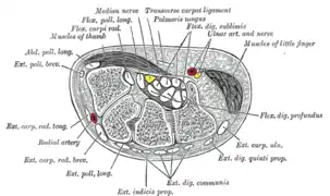 Transverse section across the wrist and digits. (Ext. carp. rad. long. labeled at center left.) | |
| Details | |
| Origin | lateral supracondylar ridge |
| Insertion | 2nd metacarpal |
| Artery | radial artery |
| Nerve | radial nerve |
| Actions | extensor at the wrist joint, abducts the hand at the wrist |
| Antagonist | Flexor carpi ulnaris muscle |
| Identifiers | |
| Latin | musculus extensor carpi radialis longus |
| TA98 | A04.6.02.040 |
| TA2 | 2497 |
| FMA | 38494 |
| Anatomical terms of muscle | |
The extensor carpi radialis longus is one of the five main muscles that control movements at the wrist.[1] This muscle is quite long, starting on the lateral side of the humerus, and attaching to the base of the second metacarpal bone (metacarpal of the index finger).
Structure
It originates from the lateral supracondylar ridge of the humerus, from the lateral intermuscular septum, and by a few fibers from the lateral epicondyle of the humerus.[2]
The fibers end at the upper third of the forearm in a flat tendon, which runs along the lateral border of the radius, beneath the abductor pollicis longus and extensor pollicis brevis; it then passes beneath the dorsal carpal ligament, where it lies in a groove on the back of the radius common to it and the extensor carpi radialis brevis, immediately behind the styloid process.
One of the three muscles of the radial forearm group, it initially lies beside the brachioradialis, but becomes mostly tendon early on. Passing between the brachioradialis and the extensor carpi radialis brevis, this tendon continues into the second tendon compartment together with the latter muscle.[2]
It is inserted into the dorsal surface of the base of the second metacarpal bone, on its radial side.[2]
Innervation
The extensor carpi radialis longus is a wrist extensor that is innervated by the radial nerve,[2][3] from spinal roots C6 and C7.[4] All other major extensor muscles in the superficial layer of the posterior compartment (the extensor digitorum, extensor carpi radialis brevis, extensor carpi ulnaris, and extensor digiti minimi) are innervated by the posterior interosseous branch of the radial nerve.
Function
As the name suggests, this muscle is an extensor at the wrist joint and travels along the radial side of the arm, so it will also abduct (radial abduction) the hand at the wrist.[2] That is, it manipulates the wrist so as to move the hand towards the thumb (i.e. abduction—away from the mid-position of the hand) and away from the palmar side (i.e. extension—increased angle between the palm and the front of the forearm).
Exercises
The muscle, like all extensors of the forearm, can be strengthened by exercise that resist its extension.
Example exercises
- Reverse wrist curls with dumbbells can be performed.
Additional images
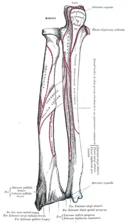 Bones of left forearm. Posterior aspect.
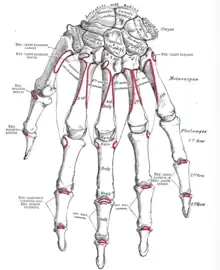 Bones of the left hand. Dorsal surface.
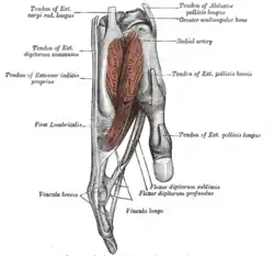 Tendons of forefinger and vincula tendina.
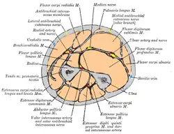 Cross-section through the middle of the forearm.
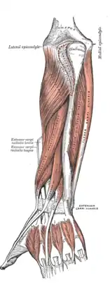 Posterior surface of the forearm. Deep muscles.
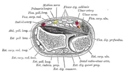 Transverse section across distal ends of radius and ulna.
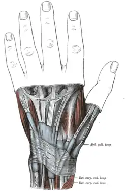 The mucous sheaths of the tendons on the back of the wrist.
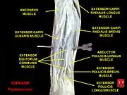 Extensor carpi radialis longus muscle
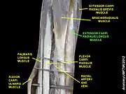 Extensor carpi radialis longus muscle
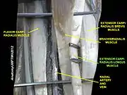 Extensor carpi radialis longus muscle
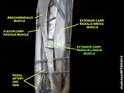 Extensor carpi radialis longus muscle
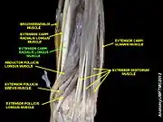 Extensor carpi radialis longus muscle
.jpg.webp) Muscles of upper limb. Cross section.
Muscles of upper limb. Cross section.
Notes
- ↑ Garten, Hans (2013-01-01), Garten, Hans (ed.), "M. extensor carpi radialis (longus and brevis)", The Muscle Test Handbook, Churchill Livingstone, pp. 58–59, doi:10.1016/b978-0-7020-3739-9.00029-8, ISBN 978-0-7020-3739-9, retrieved 2020-10-22
- 1 2 3 4 5 Color atlas and textbook of human anatomy : in three volumes. Platzer, Werner, Kahle, Werner, Frotscher, Michael (5th rev. and enlarged [English] ed.). Stuttgart: Thieme. 2003. p. 164. ISBN 978-1-58890-159-0. OCLC 54767617.
{{cite book}}: CS1 maint: date and year (link) CS1 maint: others (link) - ↑ Genet, François; Autret, Katell; Schnitzler, Alexis; Lautridou, Christine; Bernuz, Benjamin; Denormandie, Philippe; Allieu, Yves; Parratte, Bernard (December 2012). "Motor Branch of Extensor Carpi Radialis Longus: Anatomic Localization". Archives of Physical Medicine and Rehabilitation. 93 (12): 2309–2312. doi:10.1016/j.apmr.2012.03.015. ISSN 0003-9993. PMID 22459176.
- ↑ Bradley Bowden, Illustrated Atlas of the Skeletal Muscles, 2005